https://doi.org/10.22319/rmcp.v15i2.6496
Article
Prevalence of Fasciola hepatica and Calicophoron spp. in extensively reared cattle in the Florida district (Amazonas), Peru
Medali Cueva-Rodríguez a,b*
Teófilo Torrel c
Cristian Hobán d
Wuesley Alvarez-García b
Flor Mejía e
Luis Vargas-Rocha c
a Universidad Nacional Toribio Rodríguez de Mendoza de Amazonas. Laboratorio de Enfermedades Infecciosas y Parasitarias de Animales Domésticos - LABISAN, Calle Higos Urco N° 342-350-356 - Calle Universitaria N° 304. Ciudad de Chachapoyas (Amazonas) Perú.
b Instituto Nacional de Innovación Agraria. Dirección de Desarrollo Tecnológico Agrario. Estación Experimental Baños del Inca. Ciudad de Los Baños del Inca (Cajamarca), Perú.
c Universidad Nacional de Cajamarca. Facultad de Ciencias Veterinarias. Laboratorio de Parasitología Veterinaria y Enfermedades Parasitarias. Ciudad de Cajamarca, Perú.
d Universidad Nacional de Cajamarca. Facultad de Ciencias Veterinarias. Laboratorio de Inmunología. Ciudad de Cajamarca, Perú.
e Universidad Nacional Toribio Rodríguez de Mendoza de Amazonas. Instituto de Investigación de Ganadería y Biotecnología – IGBI. Ciudad de Chachapoyas (Amazonas), Perú.
* Corresponding author: mcuevar@unc.edu.pe
Abstract:
The present study determines the prevalence of eggs of Fasciola hepatica and Calicophoron spp. and of mixed infection in grazing cattle from six cattle ranches in the district of Florida, Department of Amazonas (Peru). Using the natural sedimentation technique, 358 fecal samples were examined. The prevalence of F. hepatica was 69.83 % (95% CI 65.08 - 74.59), followed by Calicophoron spp. 60.34 % (95% CI 55.27 - 65.40) and a prevalence of mixed infection 41.62 % (95% CI 36.51 - 46.73). The presence of F. hepatica eggs did not differ among farms, breeds, and age groups (P>0.05). The presence of Calicophoron spp. and mixed infection with F. hepatica showed differences between towns and breeds (P<0.05), unlike the age groups, which were statistically similar (P>0.05). A high prevalence of fecal eggs of F. hepatica and spp. was found, a situation that could be due to the environmental conditions that allow the optimal development of the intermediate host and the cattle grazing system.
Keywords: Prevalence, Coprology, Extensive breeding, Liver fluke, Rumen fluke.
Received: 23/06/2023
Accepted: 01/09/2023
Introduction
Parasitic infections are considered one of the most frequent and important health issues in grazing animals. Parasites are an obstacle to profitable livestock farming, causing reduced production and economic losses due to the costs of control, treatment, and mortality(1,2).
Fasciolosis is a disease of veterinary and public health importance that develops from the ingestion of Fasciola hepatica metacercariae in feed or drinking water(3,4). The parasite is located in the bile ducts and gallbladder, causes severe traumatic hepatitis during the migratory and biliary stages, and can lead to loss of liver function as a result of damage to liver parenchyma and bile ducts triggering liver fibrosis(5,6). In various regions worldwide, it is considered a reemerging disease and a growing threat, mainly due to the rapid evolution of human activities(7-9).
On the other hand, paramphistomosis, a disease caused by rumen trematodes of the Paramphistomidae family, has been associated with significant morbidity and severe pathological disorders such as enteritis and anemia, caused especially by the activity of juvenile trematodes in the intestine of the definitive host, the ruminant(10,11). In acute infections, the immature forms can cause the death of the animal(12). Adult parasites cause rumenitis, acute catarrhal diarrhea, hemorrhage, detachment of rumen papillae, and fibrosis, as well as the occurrence of areas with reticulum acanthosis, edema, ulceration, etc.(13-15). As in the case of F. hepatica, ruminants become infected by ingesting metacercariae encysted in forage or in water(16).
Both parasitizes are distributed across the world, mainly in tropical and subtropical regions(17,18). Because they share the same intermediate host (snails of the family Lymnaeidae), co-infections are possible in both the intermediate host and the definitive host(19). The presence of these parasites is exacerbated under favorable conditions such as wet soils, high rainfall, extensive farming systems, and fresh water bodies that host snails(20,21). On the other hand, they cause great negative economic impact on the livestock industry, affecting growth rate, feed conversion efficiency, reproductive performance, carcasses in poor condition, animals experience reductions in milk production and quality(22-25).
The lack of knowledge about the proper control of animal health issues and the low level of education of farmers, particularly in small production systems, could partly explain the high prevalence of bovine fasciolosis in certain scenarios(26). Given the frequent reports of rumen and liver parasites found in cattle slaughtered for human consumption, the importance for public health and the high economic costs involved in pharmacological treatments, the present study determines the prevalence of F. hepatica and Calicophoron spp. in grazing cattle in six annexes of the Florida district of the Department of Amazonas (in Peru). In this way, it was seek to understand and achieve a more accurate picture of the presence of both trematodes in cattle in the study area, with the consequent adoption of preventive and prophylactic measures.
Material and methods
Study area
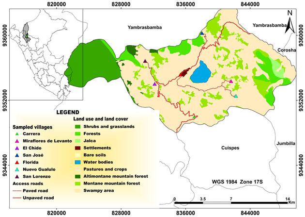
The study covered six villages in the district of Florida (Figure 1), located in the province of Bongará, Department of Amazonas, in the northeastern Peruvian Amazon. The study area has a humid tropical climate with frequent rainfall throughout the year and an average annual temperature of 16 °C.
Figure 1: Location map of the sectors in the study area. The towns are located at an altitude ranging between 2,280 and 2,750 m asl and have a relative humidity of 70 to 95 %
Animal selection and feces sampling
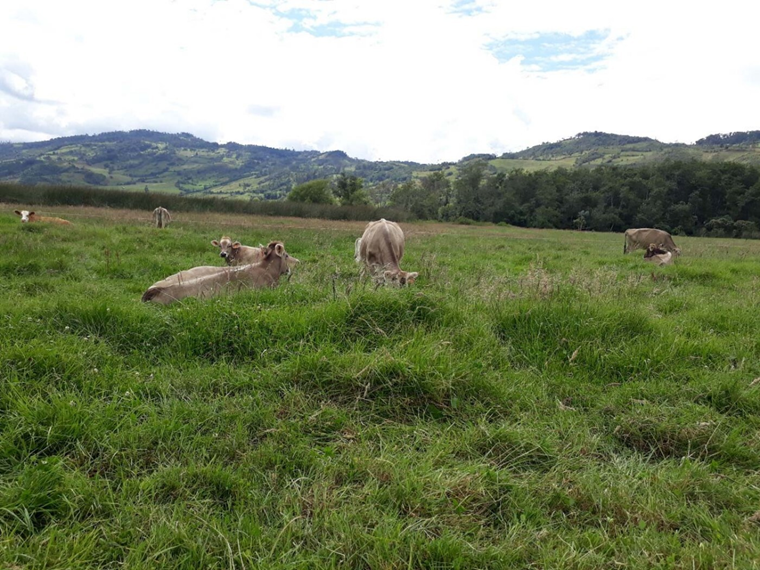
The sample size (n= 358) was estimated based on a population of 5,200 cattle (previous census), an expected proportion of 0.5, a 95 % confidence level, and a precision level of 5%. A stratified sampling with allocation proportional to the number of cattle determined the number of samples to be considered for each sector. Female cattle over 2 yr of age and of any breed were considered. Identification and age were taken from the ear tags. The animals were raised in extensive rearing systems, fed rye grass (Lolium multiflorum), clover (Trifolium repens), Kikuyu (Pennisetum clandestinum), and other native grasses (Figure 2).
Figure 2: Evaluated Brown Swiss cattle raised in open fields and fed green forage
Fecal samples (approximately 100 g) were collected directly from the rectum of the animals using sterile obstetric gloves. Each animal was restrained by the owner with the help of a rope, trying to cause the less pain as possible, with hands covered with latex gloves and the perianal region was washed with soap and water. The samples were transported to the Immunology Laboratory of the Faculty of Veterinary Sciences, National University of Cajamarca (Universidad Nacional de Cajamarca) in an expanded polystyrene box with cooling gels (2 to 4 °C). The transfer time lasted between 8 and 10 h. In the laboratory, they were kept refrigerated at 4 °C until processing after 24 h. Clean and labeled materials were used to avoid cross-contamination.
Analysis of the samples
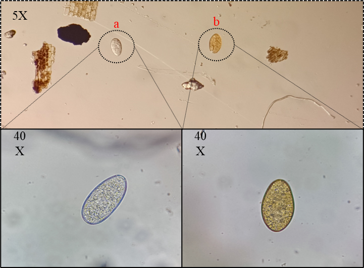
Samples were processed by natural sedimentation(27). Eggs were observed under a stereoscope with halogenated light at 5X (Nikon SMZ 745 - USA), and identification was based on the morphological characteristics of the egg of each parasite(28-31).
Cattle breeders' attitude towards parasites in cattle
According to the observations made during the fecal sample collection process, it was found that farmers in the evaluated areas lacked knowledge about mechanisms for the prevention and control of trematodes in their animals. No control or prevention measures were identified, such as the proper management of excreta, the management of drinking troughs, the implementation of drainage systems on farms, the adoption of technified irrigation practices, or the implementation of strategies aimed at controlling the intermediate host, among others. In addition, no consistent information was obtained regarding the existence of deworming programs; in fact, farmers were unaware of the presence of rumen trematodes in their animals.
According to cattle ranchers, they sometimes perform deworming with albendazole-based chemicals to control F. hepatica, known locally as Fasciola, Liver fluke, Alicuya, Lunguash, Liver slug, Dystoma, and Coca Leaf. This process was carried out during the rainy season (December to April) or when the animals presented persistent diarrhea or showed signs of decay, without the supervision of a livestock professional, and in the absence of a parasitological laboratory diagnosis by observation of fecal eggs or by any other method.
Statistical analysis
Data were processed using descriptive statistics. Positive cases were expressed as percentages with 95% confidence intervals(32). The degree of association between prevalence by sector, race, and age group was determined using the nonparametric Kruskal-Wallis test. The Phi correlation function was used to determine the degree of the linear relationship between the mixed prevalence of trematodes.
Results
Brown Swiss and Fleckvieh cows raised on pasture, ranging from 2 to more than 6 yr of age, were found in the six farms of the Florida district. Eggs of Fasciola hepatica and Calicophoron spp. were observed in all sectors (Figure 3). F. hepatica eggs were observed in 69.83 % (95% CI: 65.08-74.59) of the animals, followed by Calicophoron spp. 60.34 % (95% CI: 55.27-65.40) and a prevalence of mixed infection of 41.62 % (95% CI: 36.51-46.73) (Table 1).
Figure 3: Calicophoron spp. eggs (a) and Fasciola hepatica (b). Stereomicroscopic view (top) and microscopic view (bottom)
Discussion
The trematode Fasciola hepatica outnumbered Calicophoron spp. by less than a tenth (9.12 %), with an overall mixed infection prevalence of 41.16 % (95% CI 36.09 - 46.23). The presence of F. hepatica eggs did not differ between sectors, breeds, or age groups (P>0.05). The prevalence of Calicophoron spp. and mixed infection with F. hepatica showed differences between sectors and breeds (P<0.05), unlike the age groups, among which they were statistically similar (P>0.05). The Phi correlation showed variable results of the associated occurrence of both parasites in the animals.
The high presence of parasite eggs may be due to the good development of the intermediate host in optimal environmental conditions, humid areas, and a variety of temperatures and altitudinal levels, for example. The locations evaluated range from 11 to 20 ºC and have a relative humidity of 60 to 95 %. Since both trematodes share the same intermediate host, freshwater pulmonate mollusks of the family Lymnaeidae(33,34), this condition would facilitate co-infection in both the intermediate and definitive host.
The areas where the sampling was carried out are located between 1,300 and 2,750 m asl, ideal ranges for the development of parasitoses. Parasitic forms of F. hepatica have been reported even in intermediate hosts below 400 m asl(35) and up to 4,500 m(36). Calicophoron microbothrioides can be found below 200 m and also in mountainous areas above 3,000 m, where there is stagnant water available for the cycle of the intermediate hosts, in areas used for livestock farming, and in the areas without water for the cycle of the intermediate hosts(37).
Climate influences the rate of parasitic infection in livestock(38,39). As shown in Figure 1, the study area includes large vegetation and bodies of water, ―favorable conditions for the development of the intermediate host. In general, the province has a humid tropical climate with frequent rainfall throughout the year and an average annual temperature of 16 °C. In an area close to the study area, researchers found that water sources, mainly streams, irrigation ditches and rivers, are risk factors for F. hepatica(40).
The breed of cattle has been reported as a risk factor in several studies. Purebreds are more susceptible to infection than crossbreds(9). In a study conducted in Amazonas (Peru), it was determined that the Brown Swiss breed is more susceptible to infection by F. hepatica and other parasites(40). However, it should be noted that, in their study, the sample size for this breed was larger than for other breeds. In the present research, a higher prevalence of trematodes was found in the Fleckvieh breed. Despite an evident higher number of Brown Swiss (n= 203) versus Fleckvieh (n= 155) cattle, the prevalence was statistically equal; therefore, the results do not consolidate the breed as a risk factor.
Similarly, another study reported that the Simmental breed was a risk factor for F. hepatica infection compared to Brown Swiss and other breeds(41). Although the sample size of the Simmental breed was larger (as in the present study), the results were statistically similar to those of Brown Swiss, only differing with the Jersey, Holstein and crossbred breeds, although the sample size of these breeds was very low. Similar to the present study, several authors have not reported conclusive results in which breed is a risk factor, but rather that the presence of parasites is influenced by a higher population of a certain race in a certain place(14,42,43).
The age of the definitive host is also closely related to infection(15). As in other reports, the presence of parasites was higher in older animals. Most authors show that the prevalence of trematodes is higher in animals older than 2.5 yr(21,43,44). No association has been found between age and rumen trematode infection(14). Infected animals have an age limit for becoming infected, since the life cycle of Paramphistomidae lasts at least 6 to 8 mo(9); therefore, animals 12 to 24 mo of age may be at higher risk of infection. In addition, the animals are raised under grazing conditions from birth until they leave the herd.
Several studies have indicated that extensive animal husbandry is a factor in parasite infection(40,43). Cattle managed in extensive or semi-extensive regimes where access to pasture occurs almost year-round throughout the animal's life are more predisposed to grazing than those managed in an intensive or semi-intensive system(45).
The high prevalence of F. hepatica in cattle has been reported within the same Amazonas region, with a prevalence of 45.6 %(40), and 59.5 %(41). The presence of trematodes in cattle has also been described in other regions of Peru. In three districts of the province of Oxapampa (Pasco - Peru), by rapid sedimentation of 408 samples of dairy cattle, a prevalence of 10.0 ± 2.9 % of F. hepatica and 28.4 ± 4.4 % of a digenean of the Paramphistomidae family were found(46).
Although studies on rumen trematodes in cattle in Peru are scarce, C. microbothrioides has been reported in Amazonas(31). Trematode eggs of the Paramphistomidae family were identified in the Loreto region (Peruvian jungle)(47). In San Martin (Peruvian jungle region) Cotylophoron sp. has been reported in cattle(48). Both parasites have been identified in different parts of the world. In South America, Cotylophoron cotylophorum has been described in Colombia(49), Cotylophoron marajoensis n. sp. in Brazil(49) and C. microbothrioides in Chile(37). As well as in the Americas, the presence of both trematodes has been reported in European(50,51,52), African(34,26,38), Asiatic countries(53), etc. These regions have similar conditions to those of the present study ―extensive breeding, climatic conditions, age, breed, etc.―, as risk factors. Climate change and globalization contribute to the distribution of parasites in a territory where the intermediate host has adapted(19).
Due to the high prevalence of trematode eggs identified in the sampled areas, it is suggested that this situation may be attributed to the conditions in which cattle are raised, where no formal programs for the control and prevention of parasitoses are available. Despite the possible use of albendazole for the management of F. hepatica, no local studies of chemical-based antiparasitic efficacy have been reported.
Conclusions and implications
A high prevalence of fecal eggs of Fasciola hepatica and Calicophoron spp. was detected. Climatic and geographic conditions, in addition to the grazing system and the absence of control and prevention programs, predispose to the high presence of both trematodes. However, further studies are needed to evaluate drainage systems, pasture, and watering trough management practices in the control and prevention of trematodes, as well as evaluations of antiparasitic resistance and comprehensive studies from a One Health approach.
Sources of financing
This work was financed by the scholarship and co-financing program of the CONCYTEC, CIENCIACTIVA of the Ministry of Education of Peru (Conv-191-2015-Fondecyt). M.C.-R. is grateful to CONCYTEC for the financing. The authors are grateful to Dr. Rodrigo Sanabria, VD, researcher of the Faculty of Veterinary Sciences, UNLP – Argentina, for the guidance and direction of the research.
Conflict of interest
The authors declare that they have no conflict of interest that could have interfered with the results of this research.
Literature cited:
Table 1: Prevalence (%) of trematodes found in grazing cattle in the district of Florida, Amazon (Peru)
ab For each variable, the different letters between their levels are significant differences in each factor (Kruskall-Wallis, P<0.05).