https://doi.org/10.22319/rmcp.v15i2.6322
Technical note
Frequency of seropositivity against porcine circovirus type 2 (PCV2) in the metropolitan area of Monterrey, Nuevo León, and its peripheral area
José Pablo Villarreal-Villarreal a
César Dávila-Martínez a
Heidi Giselle Rodríguez-Ramírez a*
a Universidad Autónoma de Nuevo León. Facultad de Medicina Veterinaria y Zootecnia. Campus de Ciencias Agropecuarias, Colonia Ex-Hacienda el Canadá, General Escobedo, Nuevo León, México.
*Corresponding author: rdzmvz@gmail.com
Abstract:
Porcine circovirus type 2 (PCV2) is a DNA-type virus that has an affinity for cells of the immune system and generates lymphocyte depletion so it favors the development of diseases caused by other opportunistic agents, it is also related to the generation of different syndromes. Therefore, this virus causes major illnesses on the pig industry; nevertheless, PCV2-associated syndromes can easily be prevented through the proper application of biosecurity and vaccination measures. On the other hand, small-scale production units (SPUs) often lack this type of preventive management, as well as routine surveillance by a veterinarian. Although PCV2 is considered a widely distributed virus, there are no reports of its presence in SPUs in Nuevo León. The presence of antibodies against PCV2 was determined using a commercial kit and complete blood count was performed on the animals. A total of 48 SPUs were found, with 91.67 % positivity and 89.7 % seropositivity in the animals. In the complete blood count, it was found that HGB and HCT were decreased in individuals who were positive for antibodies compared to negative ones (P=0.03 and P=0.01, respectively); on the contrary, the value of total white blood cells was found to be decreased in individuals who were negative for the presence of antibodies against PCV2 (P=0.01).
Keywords: Backyard, PCV2, Porcine circovirus type 2, ELISA, Seroprevalence, Pigs.
Received: 13/03/2023
Accepted: 19/01/2024
Porcine circovirus type 2 (PCV2) is a single-stranded DNA virus that only infects pigs, so it has no zoonotic significance. PCV2 is a pathogen that is implicated in the development of different syndromes, such as post-weaning wasting syndrome, dermatitis and nephropathy syndrome, as well as reproductive failure(1). It is recognized that infection with this virus is a predisposing factor for syndromes, however, it requires co-infection with another pathogen to trigger the disease process. Among the risk factors that increase the possibility of introducing the agent into the pig population and its dissemination are: low birth weight, low weaning weight, as well as those related to facilities and management practices, such as housing a large number of animals in a small space, contact between pigs and hygiene(2).
PCV2 has an affinity for the cells of the immune system, especially macrophages and lymphocytes(3); in fact, one of the expected clinical findings during infection by this virus is the reduction of total leukocyte counts, as well as lymphopenia(1,4,5). Among the diagnostic methods for this virus are PCR and immunohistochemistry (IHC)(6); for the latter, lymphatic tissue is used to detect antigens of the virus, which indicates the presence of the virus in the target cells. It has been confirmed that lymphocyte depletion in lymphatic tissue is related to the activation of apoptosis through the pathways of caspases 3 and 8 within lymphocytes(3), although it is not ruled out that there are other mechanisms involved. Lymphocyte depletion induces a state of immunosuppression that is also aggravated by co-infection with other pathogens, such as porcine parvovirus, PRRS virus, and others(7), which allows the appearance of the aforementioned syndromes.
In Mexico, small-scale production units (SPUs) are still present in some areas. The characteristics of these systems present in the metropolitan area of Monterrey, Nuevo León, have previously been researched, and among them, the lack of biosecurity, medication and routine surveillance by a veterinarian has been confirmed. The aforementioned conditions favor the entry of pathogens into production units, as well as their subsequent perpetuation in the environment; nevertheless, PCV2 is especially important since being immunosuppressive, it allows the appearance of clinical manifestations of secondary infections in some cases(8-10). Therefore, this study aimed to determine the presence of antibodies against PCV2 in backyard pigs that did not have previous vaccination against this pathogen, so a cross-sectional study was carried out, which included pigs in SPUs from 9 municipalities corresponding to the metropolitan area of Monterrey, Nuevo León.
The small-scale production system (also known as backyard or artisanal) was defined as one in which pig rearing activities will be carried out in the home of the owners of the animals, or as a complementary economic activity, that is, one that will not represent the main family income. The minimum number of samples was calculated with the WinEpi software under the following considerations: an expected prevalence of 92 % was considered, which was taken from a previous report in Mexico(11), a margin of error of 5 %, and a confidence level of 95 % for an unknown population, yielding a minimum number of 114 samples. Once the SPUs were identified, permission was requested from the owners to take samples from the animals and a questionnaire was applied at the end. Samples were collected from May 2019 to March 2020. Pigs of different ages were included; however, sampling in pregnant females was avoided to prevent the risk of abortion, as well as in suckling piglets to avoid detection of maternal antibodies.
For sampling, the pigs were physically restrained. During immobilization, the body condition of each individual was rated on a scale of 1 to 5. Two blood samples were taken from the jugular vein, one in a collection tube with EDTA and the other in a collection tube with serum separator. The samples with EDTA were processed in the clinical laboratory of the Veterinary Hospital of Small Species (HVPE, for its acronym in Spanish) of the Autonomous University of Nuevo León under the standard procedure on the KONTROLab 5R+Vet equipment.
The serum was separated from the clot at 1,000 rpm for 10 min at 4 °C and then fractionated into 500 μL aliquots and stored at -80 °C until later use. For the detection of antibodies against PCV2, a commercial kit (Bio Check®) was used according to the manufacturer’s instructions. This kit has a sensitivity of 92.1 % and a specificity of 95.6 %. The absorbance of the samples was read at 405 nm on the Awareness technology Chromate® equipment (Awareness technology Inc.).
Data were captured in a spreadsheet to determine the percentage of seropositivity. A Student’s T-test was performed to determine the difference between the means of the hematological parameters between the antibody-positive and antibody-negative groups, as well as of the frequency of the animals’ body condition. Statistical analysis was performed using the GraphPad Prism 6 software (San Diego, CA). A P-value ≤ 0.05 was considered significant.
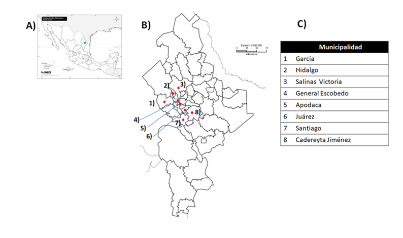
There were localized 48 SPUs, in which access to sample was allowed, which were in the municipalities of Apodaca, Cadereyta Jiménez, García, General Escobedo, Hidalgo, Juárez, Santiago and Salinas Victoria. It was found that, in 44 of the sites sampled, at least one animal tested positive for antibodies, so the percentage of positivity was 91.67 %. The rest of the production units corresponded to four sites where no animal was detected as positive for antibodies against PCV2. In all municipalities, it was possible to detect positive production units.
A total of 204 animals were sampled, of which the presence of antibodies against PCV2 was confirmed in 183, resulting in 89.7 % seropositivity. Some studies have previously been carried out in Mexico, where 92.29 % positivity for antibodies against PCV2 was determined among animals and 98.14 % positivity among production units with at least one positive animal(11), so, in congruence with findings of other authors, seropositivity against this virus was found ubiquitously in the SPUs in the metropolitan area of Monterrey, Nuevo León, and its peripheral area.
Figure 1: A) Map of Mexico. B) Map of Nuevo León. Each red dot represents a sampled municipality. C) Identification of sampled municipalities
On the other hand, different research groups have explored the presence of PCV2 through the use of real-time PCR; an example is in Brazil where they have found the presence of the virus genome in 15.6 %(8) of the lung samples studied. On the other hand, by using quantitative PCR, a 90 % prevalence has been detected in Colombia by using white blood cell samples(9). In Spain, another group of researchers have taken on the task of identifying the presence of the virus in technified production units in different areas using quantitative PCR in environmental samples, which included swabs from the surfaces of pens, workers’ boots and even inside the offices of five production units(12), finding a 42.9 % positivity rate, thus reiterating the easy spread of this virus and its wide dissemination in the environment.
The seroprevalence results obtained were entered into the WinEpi platform, and the sensitivity and specificity specifications provided by the kit manufacturer were also entered in order to estimate the positive and negative predictive values. The platform yielded a positive predictive value of 99.9 % and a negative predictive value of 25.4 %, as well as an actual prevalence of 97.3 %.
In this study, pregnant females were not sampled due to the risk of inducing abortions or, where applicable, preterm births. Nonetheless, there are studies in the context of other infectious agents, such as influenza A, in which it has been shown that females with a higher number of births have a greater immune experience due to their age, as well as a higher concentration of specific antibodies(13). Therefore, it is to be assumed that, if positivity was found in animals of other ages, there is also seropositivity among females. On the other hand, pigs are animals that are born agammaglobulinemic unless they are exposed to an agent in utero, therefore, the intake of colostrum is important for their survival, since in this way they acquire IgG and IgA from the mother(14); in this study, piglets that had not been weaned were not included because the presence of maternal antibodies can be detected through the ELISA method without representing seroconversion due to exposure to the agent.
Regarding the body condition of the animals, only one animal with condition of 1 (0.41 %), 49 animals with condition of 2 (20.16 %), 86 with condition of 3 (35.39 %) and 107 animals with condition of 4 (44.03 %) were observed. No animals with a condition of 5 were observed. Mean body condition in the antibody-positive group was compared with the negative group (n= 139 animals), but no difference was found (P>0.05). Since vaccination against PCV2 was not reported for any of the animals, it is presumed that the presence of antibodies is due to seroconversion due to previous immune experience against the field virus, however, these antibodies could be fulfilling a protective role against the development of PCV2-associated syndromes. It has been shown that vaccination does not always prevent viremia, but it does reduce systemic viral load in vaccinated individuals(15). In addition to the above, a group of researchers demonstrated that vaccination has a positive effect on the cellular and humoral immune response even in animals that previously had viremia(16), that is, that had been infected before being vaccinated.
Although a high frequency of antibody positivity was found in this study, no animals with apparent clinical symptoms were found. Most of the animals had medium to good body condition; it was not possible to differentiate antibody-negative animals from positive ones by body condition either. Other researchers have found that the presence of the virus on farm is not necessarily compatible with the presence of PCV2-associated syndromes(12), and although this virus is recognized as necessary to trigger associated syndromes, its presence alone is not sufficient to produce the disease(17).
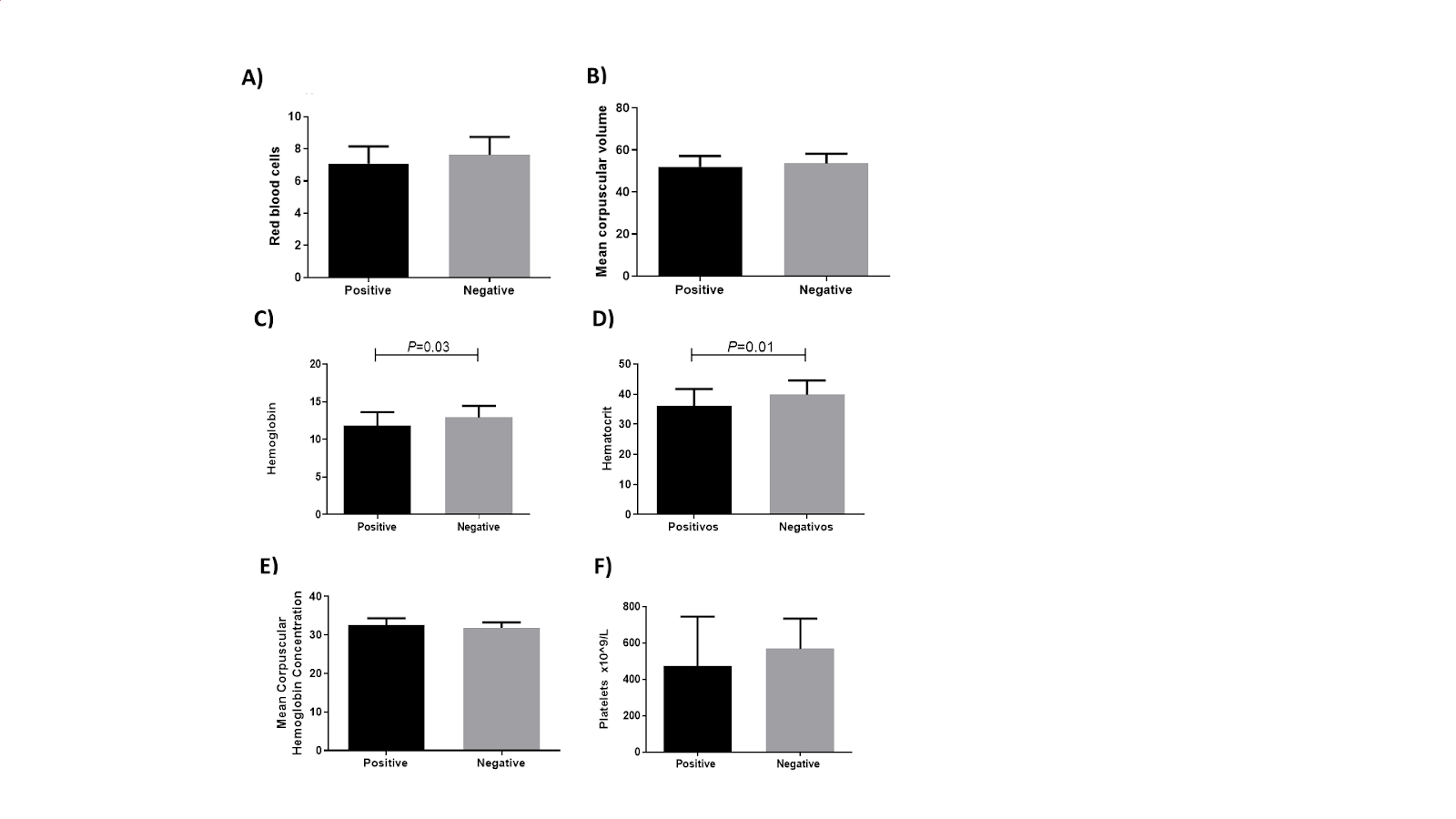
On the other hand, the means of the hematological parameters in the antibody-positive group were compared with the negative group, and in terms of the red line, a significant difference was found for hemoglobin (HGB) (P=0.03) and hematocrit (HCT) (P=0.01), which were found to be decreased in the positive group compared to the antibody-negative group (Figure 2).
Figure 2: Comparison of hematological parameters in the red line
The number of individuals included in each analysis were: A= 124, B= 124, C= 141, D= 141, E= 141, F= 141.
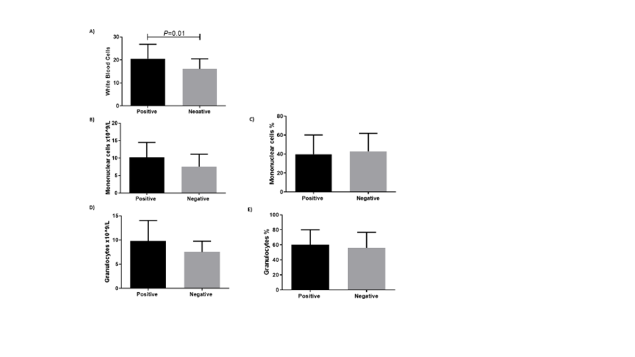
For the white line (Figure 3), a decrease in total white blood cells was found in the group negative for antibodies against PCV2 compared to those who were seropositive (P=0.01). Although there were found differences in the decreased parameters of HGB and HCT in the positive individuals and total white blood cells in greater amounts in the antibody-positive compared to the negative ones, all three parameters were within the expected normal ranges in both groups. Interestingly, it was found a decrease in the total leukocyte count in the antibody-negative group; nevertheless, the finding suggests that these pigs could be in a state of infection and even viremia in which they have yet to develop antibodies; likewise, it must be taken into consideration that most of the individuals who remained negative for the presence of antibodies were in places where at least one positive animal was found, so it is very likely that they will have contact with the virus at some point in their lives.
Figure 3: Comparison of hematological parameters in the white line
The number of individuals included in each analysis were: A= 147, B=81, C=134, D= 81, E= 134.
In conclusion, there is presence of antibodies against PCV2 in a large proportion of the SPUs and in the pigs within them, which are located in the metropolitan area of Monterrey, Nuevo León, and its peripheral area; however, this does not imply the presence of clinical symptoms.
Acknowledgements
We are grateful for the financial support for the research provided by the Autonomous University of Nuevo León in the PAICyT 2021 call, to carry out this project.
Conflicts of interest
The authors declare that there is no conflict of interest.
Literature cited: